ㄴㄴ
J Neurosonol Neuroimag Search

CLOSE


J Neurosonol Neuroimag > Volume 15(2); 2023 > Article
Morais, Mendes, Verde, Lucena, Mont’Alverne, Lima, and Carvalho: The Music Within: A Doppler Harmonic Pattern Report in Non-Subarachnoid Hemorrhage Related Cerebral Vasospasm

Abstract

A 40-year-old male presented an episode of severe thunderclap headache with right hemiparesis. Imaging revealed severe stenosis in the left middle cerebral artery, associated with increased mean velocities detected by Transcranial Doppler Ultrasound (TCD). Notably, a distinctive "tin whistle" sound, known as a doppler harmonic effect, near the highest velocity point was observed during the examination. Extensive workup was performed, including CSF analysis and laboratory investigations, and the diagnosis of Reversible Cerebral Vasoconstriction Syndrome (RCVS) was established. Treatment involving medication and daily monitoring led to a decline in the left MCA's velocity, enabling the patient's discharge with no recurrent headaches or neurological deficits. Notably, a follow-up MRI exhibited the disappearance of the left MCA stenosis. This report discusses the rare occurrence of "musical murmurs" in cerebrovascular diseases, underscoring the potential for early diagnosis using techniques like TCD in emergency settings, particularly in conditions like RCVs.

Doppler harmonic patterns were initially described in the examination of cardiac structures, mainly stenotic valves, as a manifestation of turbulent flow through a narrow space.1 The so-called musical murmur, in its pure tone quality, is a rare presentation in cerebrovascular diseases. They are often associated with subarachnoid hemorrhage (SAH), and to our knowledge, have never been documented in association with reversible cerebral vasoconstriction syndrome (RCVS).2,3

CASE

A 40-year-old male presented to the neurological Emergency Department with a history of severe left-sided headache four days earlier. He was evaluated at a secondary care facility on the first day but was discharged home a few hours later after a normal computed tomography (CT) scan and with the prescription of symptomatic medications. Twenty-four hours later he referred a new episode of a thunderclap headache associated with right hemiparesis which resolved within 40 minutes and was admitted to our facility presenting only slight right hypoesthesia.
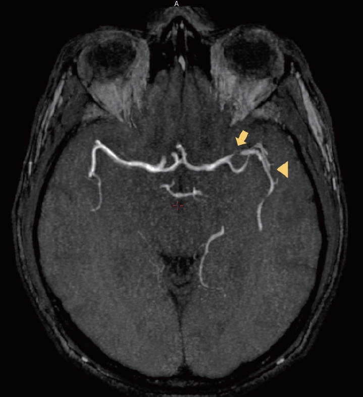
The admission CT scan revealed no signs of bleeding or ischemia. Cerebrospinal fluid (CSF) analysis revealed normal cytology, protein, glucose, and negative microbiology (including gram stain, fungi and acid-fast bacilli) and also negative venereal disease research laboratory test (VDRL) and fluorescent treponemal antibody absorption test (FTA-ABS). The patient underwent an extensive diagnostic workup including the following tests: antinuclear antibodies (ANA), VDRL, Chagas Disease, HIV, B and C Hepatitis. His erythrocyte sedimentation rate (ESR) was of 12 mm and C-reactive protein (CRP) was 1.99 mg/L. An magnetic resonance (MR) angiography showed severe stenosis of the M1 and distal M2 segments of left middle cerebral artery (MCA). A less severe stenosis of the right M1 segment of right MCA is also seen (Fig. 1).
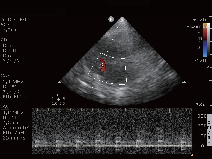
Transcranial doppler ultrasound (TCD) was also performed, revealing increased mean velocity at both MCAs (116 cm/s on the right and 181 cm/s on the left). During the examination, a “tin whistle” or sound with spectral harmonic representation was identified close to the highest velocity rate sign of the left MCA (Supplementary Video 1; Fig. 3). The patient was treated with analgesics, oral nimodipine, IV fluids, and daily TCD monitoring. A progressive decline of left MCA mean velocity was observed since admission. After 16 days, the mean left MCA velocity was 126 cm/s and he was discharged home with no headache recurrence or neurological deficits.
Diagnosis of RCVS was made based on clinical criteria associated with complementary CSF, laboratory tests and image analysis.4 He remained with no deficits. A Follow-up magnetic resonance angiography (MRA) was performed 10 months later and revealed disappearance of the left MCA stenosis (with only a slight irregularity of the left MCA-M1 segment considered not clinically significant) (Fig. 2).

DISCUSSION

Musical murmurs (MM), often described as “seagull or goose cry”, are physical phenomena of harmonic sound reverberation between two nodes when blood flows through a narrow space. They are represented on Doppler waveforms as strings oscillations that mirror each other (Fig. 3). 3,5 The turbulence generated by this kind of flow is frequently described as a result of vortex shedding, such as Kármán vortex street. Its dynamic is manifested in bodily fluids, wind flowing, and even channelized to create resonance as an ancient musical instrument like the aeolian harp.2,6
MM are still considered a rare cerebrovascular disease manifestation (0.5%) and are related to severe stenotic patterns mainly in intracranial vessels.3 Case reports were described in association with cerebral vasospasm due to SAH, arteriovenous malformation, and carotid dissection.7,8 To our knowledge, this is the first case report of musical murmur described in a patient with RCVS. TCD is still underused in the emergency setting and can be particularly useful in situations where intracranial stenosis is suspected. MM may not be a rare occurrence, since it is not promptly recognized. Identifying this phenomenon at the emergency department may lead to an earlier diagnosis of critical stenosis and improved follow-up such as in RCVS.

SUPPLEMENTARY MATERIALS

Supplementary materials related to this article can be found online at https://doi.org/10.31728/jnn.2023.00141.
Supplementary Video 1.
“Tin whistle” or “seagull/goose cry” musical sound characteristic of severe stenosis.

NOTES

Ethics Statement
This report was approved by the institution's ethics committee under CAAE number 70426623.0.0000.5040.
A written consent from the patient was obtained.
Availability of Data and Material
All data related to this study are included in the main text.
Sources of Funding
None.
Conflicts of Interest
No potential conflicts of interest relevant to this article was reported.

Acknowledgments

This publication was only possible due to efforts of residents and staff team to provide science and groundbreaking knowledge to the formation of neurologists meanwhile providing the best treatment possible for brazilian public health system (SUS) users.

Fig. 1.
Angio-MRI displaying severe stenosis of left cerebral medium artery (CMA) (arrow). Further stenosis after bifurcation of CMA (arrow head). MRI, magnetic resonance imaging.
jnn-2023-00141f1.jpg
Fig. 2.
Angio-MRI displaying resolution of the left and right middle cerebral artery (MCA) stenosis (a non-clinically significant irregularity of the left MCA-M1 segment is seen). MRI, magnetic resonance imaging.
jnn-2023-00141f2.jpg
Fig. 3.
TCD displaying harmonic pattern wave. TCD, transcranial doppler ultrasound.
jnn-2023-00141f3.jpg

REFERENCES

1. Stein PD, Sabbah HN, Lakier JB. Origin and clinical relevance of musical murmurs. Int J Cardiol. 1983;4:103-112.
crossref pmid
2. Aaslid R, Nornes H. Musical murmurs in human cerebral arteries after subarachnoid hemorrhage. J Neurosurg. 1984;60:32-36.
crossref pmid
3. Lin SK, Ryu SJ, Chang YJ, Lee TH. Clinical relevance of musical murmurs in color-coded carotid and transcranial duplex sonographies. AJNR Am J Neuroradiol. 2006;27:1493-1497.
pmid pmc
4. Rocha EA, Topcuoglu MA, Silva GS, Singhal AB. RCVS2 score and diagnostic approach for reversible cerebral vasoconstriction syndrome. Neurology. 2019;92:e639-e647.
pmid
5. Valdueza JM, Schreiber SJ, Roehl JE, Connolly F, Klingebiel R. Neurosonology and neuroimaging of stroke: a comprehensive reference. 2nd ed. Stuttgart: Thieme; 2017.

6. Britannica, The Editors of Encyclopaedia. Aeolian harp. Encyclopedia Britannica. 2019 [cited 2022 Oct 21]. Available from https://www.britannica.com/art/Aeolian-harp.

7. Morelli N, Immovilli P, Mazza L, Rota E, Spallazzi M, Rocca G, et al. “The seagull cry” in internal carotid artery dissection. Neurology. 2011;77:1769.
crossref pmid
8. Morelli N, Rota E, Michieletti E, Guidetti D. The “seagull cry” in a patient with a cerebral arteriovenous malformation. J Ultrasound. 2012;15:176-178.
crossref pmid pmc


ABOUT
ARTICLE CATEGORY

Browse all articles >

BROWSE ARTICLES
AUTHOR INFORMATION
Editorial Office
1412, 359, Gangnam-daero, Seocho-gu, Seoul, Republic of Korea
E-mail: ksn@neurosonology.or.kr                

Copyright © 2024 by Korean Society of Neurosonology.

Developed in M2PI

Close layer
prev next