ㄴㄴ
J Neurosonol Neuroimag Search

CLOSE


J Neurosonol Neuroimag > Volume 15(1); 2023 > Article
Walter: Muscle Ultrasound in Clinical Neurology: Diagnostic Uses and Guidance of Botulinum Toxin Injection

Abstract

Muscle ultrasound (MUS) is increasingly used by neurologists, neuropediatricians, neurosurgeons, specialized radiologists and anaesthesiologists for the imaging-supported diagnosis of neuromuscular disorders. Especially, MUS is highly sensitive in detecting fasciculations in motor neuron diseases, and in revealing intensive care unit acquired weakness. Hereditary and inflammatory myopathies are associated with distinct patters of echo-intensity changes of affected muscles. Moreover, MUS can be used for guiding needle biopsy of muscle lesions, and for targeting intramuscular botulinum neurotoxin (BoNT) injection in neurological disorders with muscle hyperactivity. MUS-guidance of BoNT injection is especially recommendable in complex cervical dystonia, in task-related hand dystonia (writer’s cramp, musician dystonia), and in children and adolescents with cerebral palsy. Modern ultrasound technologies such as sono-elastography, tissue Doppler, and high-definition microvasculature imaging allow for novel diagnostic and therapeutic uses. Recently, an international expert group reported consensus guidelines for neuromuscular ultrasound training. The present review provides a concise overview of well-established diagnostic and therapeutic applications of MUS in clinical neurology, with specific focus at MUS for targeting intramuscular BoNT injections.

INTRODUCTION

Muscle ultrasound (MUS), also referred to as myosonography, has been applied in the field of neurology since the early 1980s, even though ultrasound (US) image resolution was low at that time.1-4 With the advance of imaging technology MUS became more feasible, and meanwhile numerous diagnostic and therapeutic applications have been developed.5-8 In 2019, an international expert group reported consensus guidelines for neuromuscular US training including both, assessment of nerves and muscles.9 According to these guidelines the following procedural skills are required specifically for MUS: basic level—(1) differentiating bone, muscle, nerve, vasculature, and tendons; and (2) discrimination of normal vs. abnormal muscle (e.g., Heckmatt scale, atrophy, echogenicity, and thickness measurement); intermediate level—(1) patterns of muscle involvement in generalized neuropathy/myopathy; and (2) dynamic muscle ultrasound (e.g., fasciculations/diaphragm excursion); advanced level—(1) quantifying muscle echogenicity; (2) needle guidance for intramuscular injections (e.g., botulinum toxin); (3) needle guidance for muscles with increased risk (e.g., diaphragm); and (4) advanced MUS techniques, such as elastography, 3D, extended field of view, and contrast agents. The present review provides an overview of well-established applications MUS in clinical neurology, with specific focus at MUS for targeting intramuscular botulinum neurotoxin (BoNT) injections. This article follows the outline of the author’s lecture held on November 4, 2022, during the second day of the Joint Conference of the Korean Society of Neurosonology (KSN) and the Neurosonology Research Group of the World Federation of Neurology (NSRG-WFN) in Seoul, Korea.

TECHNICAL CONSIDERATIONS

US machines used for MUS are usually identical to those used for vascular US. In principle, all contemporary US machines can be used if equipped with an appropriate linear-array or, alternatively for deep muscles, a curved-array transducer. The applied US frequency determines both, the image resolution and the penetration depth. Increasing frequencies increase image resolution, but reduce penetration depth and vice versa. Profound and large target muscles in the neck, trunk and leg are best visualised with US frequencies of about 7.5 (5–10) MHz.10 The assessment or targeting of superficial and small muscles in the face or arm region requires optimal resolution and therefore US frequencies in the high-frequency range of about 18.0 (15–25) MHz (Table 1).6,11,12 For standard applications of B-mode MUS, the US system settings are adapted at high dynamic range and at medium-to-high persistence. Contemporary US systems are usually delivered by the manufacturer with a choice of ‘musculoskeletal’ presets that can be selected for MUS.6 For optimal image quality a rectangular probe position with only moderate pressure on the underlying tissue is recommended. Angular deviation of the probe position may reduce tissue echogenicity, increased pressure of the probe towards the skin may increase tissue echogenicity. Rectangular probe position in relation to bone surfaces is recognized by the very bright appearance of the surface of the referring bone. To allow for improved comparability it has been recommended that the left side of the US screen shows the medial, ulnar, cranial or proximal side of the examined body part and the right side the lateral, radial, caudal and distal side.13 Alternatively, at axial transection of a body part the left side of the US image can display the left side of the referring body region as seen from the investigator which may be more intuitive especially if MUS is applied for injection guidance.6

ULTRASONOGRAPHY OF NORMAL MUSCLE

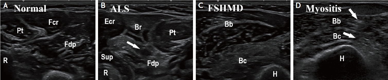
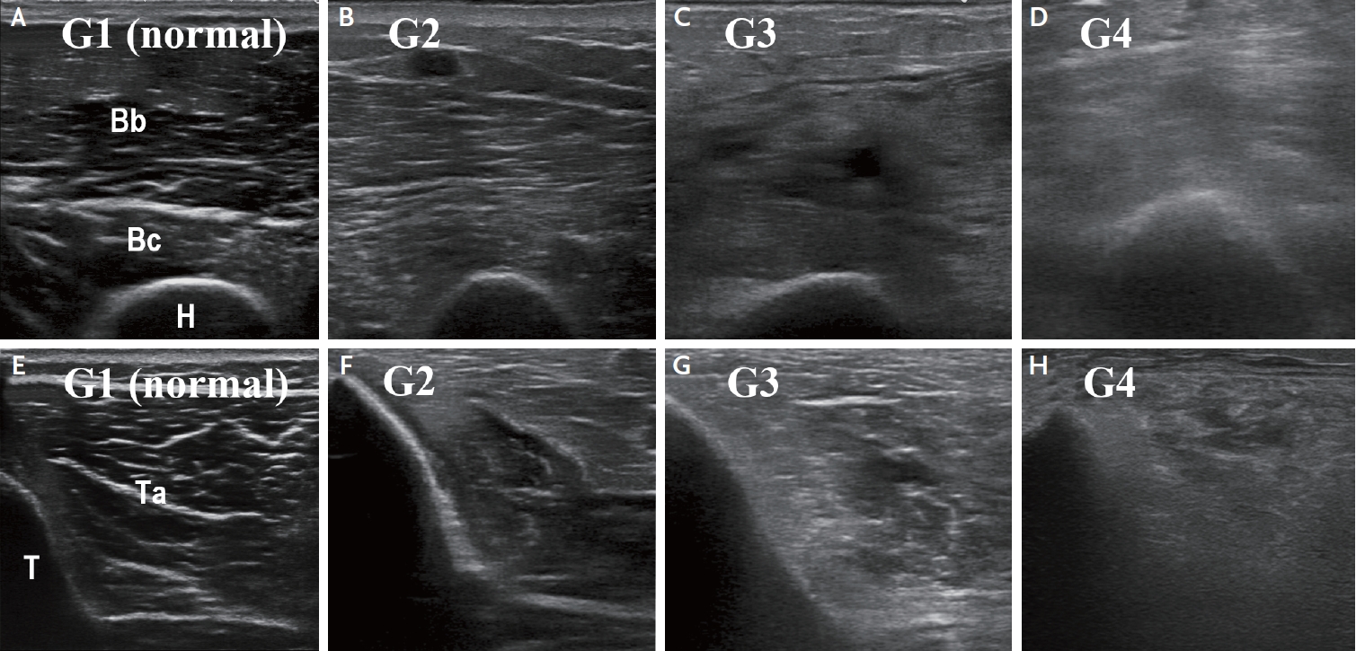
Normal muscles can be identified easily on US by their “starry night” appearance on transverse view and pennate appearance on longitudinal view, the identification of their origin and insertion and their diameter changes on contraction.10,14 Muscle tissue has normally a low echogenicity. Its echogenicity is increased in the presence of fibrosis and by infiltration of fat tissue which may be caused by a number of neuromuscular disorders (see below).3-5,15 Sizes of human limb muscles, paraspinal muscles and even of cranial or facial muscles can be reliably measured with US.16-19 Individual muscle fibres which are striated, multinucleated cells, ranging from 10–100 µm in diameter, can be distinguished from each other with ultrahigh-frequency (>30 MHz) MUS,20 but not with standard MUS. Muscle fibres are organized in grouped bundles (fascicles) surrounded by fibroadipose septa called perimysium. Hyperechogenic perimysium can be distinguished from muscle fascicles with diameters of more than 2 mm. The highly echogenic epimysium allows distinguishing muscles from each other.10 Figs. 1A, 2A, 2E, and 3E show MUS images of normal muscle tissue surrounded by other tissues.

HEREDITARY AND INFLAMMATORY MYOPATHIES

Myopathies with degeneration of muscle fibers are characterized by changes of tissue composition in the affected muscles. Damaged muscle fibres are replaced with connective and fat tissue which results in increased muscle echo-intensity.21 Increased muscle echogenicity is most pronounced in chronic injury due to muscular dystrophy, myositis, or nerve injury without re-innervation (Fig. 1).22-24 Both, the echotexture pattern within an altered muscle and the pattern of involved muscles on MUS, can aid the differentiation of muscle disorders (Table 2). The increase of echo-intensity can be graded visually (qualitative MUS) or by use of a digitized image analysis software tool (quantitative MUS). For visual grading the most widely used scale is the Heckmatt scale (Table 3, Fig. 2).25 For digitized analysis MATLAB based software or the standard histogram function of Adobe Photoshop (Adobe systems Inc., San Jose, CA, USA) has been applied.26,27 Qualitative and quantitative assessment of muscle echo-intensity allowed for the detection of disease progression in Duchenne and facioscapulohumeral muscular dystrophy.26-28 Qualitative and quantitative MUS detected early changes of affected muscles in children with Duchenne muscular dystrophy prior to clinical motor regression.28 Regarding inclusion body myositis (IBM), quantitative MUS (echo-intensity) of the flexor digitorum profundus muscle and the gastrocnemius muscle discriminated well between IBM patients and healthy controls, unlike MUS measures of muscle thickness.29 Beside echo-intensity and muscle thickness, being assessed on B-mode images, advanced MUS using shear wave elastography has been reported to improve the detection of myopathies such as polymyositis and dermatomyositis.30 MUS can help determine the optimal muscle biopsy site (e.g., by placing a skin mark prior to biopsy), or can be used for real-time visualization and guidance of needle biopsy.23

INTENSIVE CARE UNIT ACQUIRED WEAKNESS

Critically ill patients commonly suffer from extensive skeletal muscle atrophy and a generalized reduction of muscle strength, a condition termed intensive care unit acquired weakness (ICUAW).31 ICUAW is triggered by critical illness, immobilization and prolonged mechanical ventilation. Typically the upper and lower extremities present with flaccid, symmetrical paresis while facial muscles and cranial nerves are spared. The pathophysiological correlate comprises critical illness myopathy and critical illness polyneuropathy, with both components present in 30–50% of patients, and pure myopathy in the majority of the other patients, while pure neuropathy is rare.32 Since specific diagnostic (e.g., laboratory) markers are not established yet, the diagnosis is based mainly on clinical examinations. However, due to prolonged sedation, delirium and a cognitive impairment, clinical assessment is often limited, if not impossible in critically ill patients.33 Fortunately, an increasing number of studies demonstrated the value of MUS in the diagnosis of ICUAW.34-41 Many studies assessed echo-intensity with quantitative or qualitative MUS,34,35,37-39,42 others employed measures of muscle thickness or pennation angle.36,40,41 Increased echo-intensity of muscle tissue in ICUAW patients has been related to inflammation-induced myofiber necrosis which occurred in about half of patients within a week of ventilation on ICU.43 Data of pilot studies suggest that MUS may be helpful for the outcome prediction of patients with ICUAW.36,39 Qualitative MUS (Fig. 2) might possibly be more useful compared to quantitative MUS in diagnosing ICUAW.39,40,42 Assessment of echo-intensity of arm muscles may be more valid than MUS of leg muscles in discriminating patients who develop ICUAW from those who do not.38,39 More recently, also shear wave elastography (SWE), superb microvascular imaging (SMI), and contrast-enhanced ultrasound (CEUS) were reported to add diagnostic value in ICUAW.44 If echo-intensity is used as a diagnostic marker of ICUAW, based on the current evidence the sum score of Heckmatt grades of the following muscles (each assessed bilaterally) should be calculated: biceps brachii, brachioradialis, forearm extensors, quadriceps femoris and anterior tibial muscle.34,35,38,39

PERIPHERAL NEUROPATHIES

In patients with chronic disease of peripheral nerves, typically atrophy of the denervated muscles occurs which can be measured with MUS.22,23,45 In classical distal-symmetric polyneuropathy, atrophy is most pronounced in distal muscles.45 The denervation causes degeneration of muscle fibres which are replaced with connective and fat tissue. This results in streaky, patchy increased echogenicity interspersed with areas of relatively spared muscle with normal, low echo-intensity (moth-eaten appearance; Fig. 1B).22,23

MOTOR NEURON DISEASES

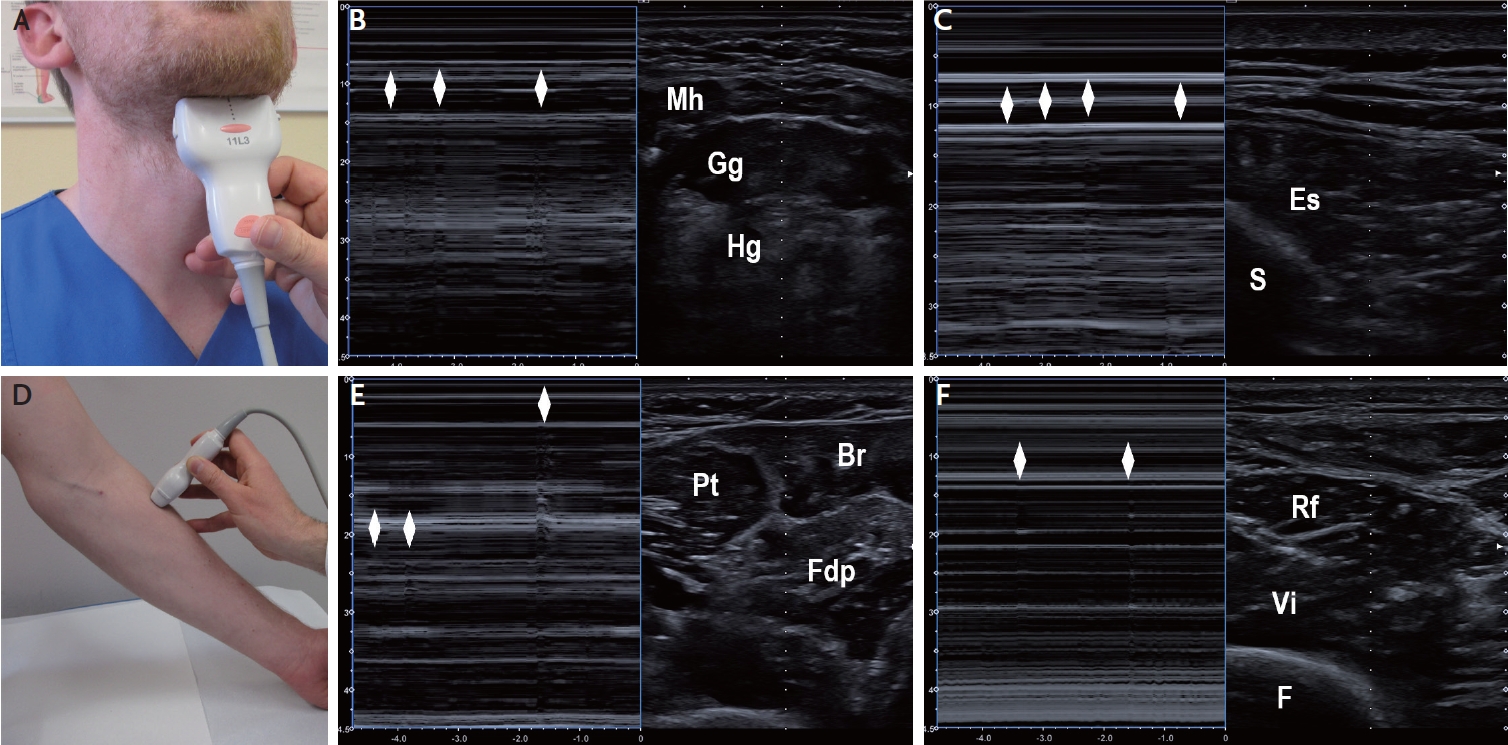
Similar to the changes in peripheral neuropathies, the denervation of muscles occurring in amyotrophic lateral sclerosis (ALS) or spinal muscular atrophy (SMA) causes measurable atrophy, increase of echogenicity and moth-eaten patterns on MUS.22,23 Histologically, muscle fibrosis is the correlate of increased echo-intensity in ALS.46 In motor neuron diseases MUS especially of the diaphragm is of diagnostic and prognostic relevance since MUS allows assessment of diaphragm function and thickness.47-49 The early affection of the abductor pollicis brevis and first dorsal interosseous muscles in ALS, with relative sparing of the abductor digiti minimi, known as the split hand phenomenon, can well be documented as the split hand index (ratio) calculated from the echo-intensities of medial versus lateral hand muscles.50 A prominent diagnostic feature mainly of ALS, but also SMA, are fasciculations in the skeletal muscles affected by the progressive denervation.48,51 Fasciculations are assessed on B-mode MUS in combination with the M-mode (motion mode) function (Fig. 3). Is has been shown that MUS is superior to electromyography in detecting fasciculations in ALS.52 The highest frequencies of fasciculations have been reported for the following muscles: biceps brachii, extensor digitorum communis, abductor pollicis brevis, first dorsal interosseous, rectus femoris, vastus lateralis, and tibialis anterior, irrespective of the region of ALS onset.52-55 Scan time per muscle of ≥60 s increases the chance of detecting at least two fasciculations per affected muscle to more than 90%.54 To detect fasciculation in the tongue, MUS is usually performed with submandibular position of the probe (Fig. 3A), however, higher sensitivity has been reported with transoral MUS.56 The diagnosis of ALS can be established if fasciculations (regarded as signs of the 2nd motor neuron) are present in two different body regions, and if additionally signs of the 1st motor neuron are present.57 SMA (and ALS) can be discriminated from hereditary muscular disorder with high sensitivity and specificity, if fasciculations are present on MUS in at least two different muscles groups.51

ULTRASOUND GUIDANCE OF BOTULINUM NEUROTOXIN INJECTIONS

1. Technique

Optimal positioning of patients, physician, and MUS device should be secured when MUS-guided injection is intended.58 Accurate clinical investigation of the patients, skilful planning of BoNT dose and injection targets, and precise anatomic knowledge are prerequisites of treatment success.6,59-64 MUS is useful not only for targeting and documentation of the injection procedure but may also aid in the identification of hypertonic (spastic, dystonic) muscles, e.g., by comparing muscle thickness and echogenicity between affected and unaffected side,60,65 or by applying sono-elastography.66-68 It has been demonstrated that MUS guidance clearly improves the precision of needle placement, especially in small and deeply located muscles.69-72 In addition MUS avoids needle puncture-related nerve or vessel injury.6,72,73 BoNT injections are usually performed with 20-mm, 27-gauge (outer diameter: 0.40 mm) needles or 40-mm, 27-gauge needles.6 For profound muscles an 80-mm, 23-gauge (0.60 mm) needle or a 120-mm, 25-gauge (0.50 mm) spinal needle may be used. Disinfection of skin and ultrasound transducer should be performed with non-alcoholic agents since alcohol may harm the transducer surface. The target muscle is first identified on MUS, and the optimal probe position for injection guidance is explored. Then the injection needle is inserted adjacent to the ultrasound transducer. While insertion of the needle through regular ultrasound gel has been proposed to be safe, it may be preferable to remove the excess gel with a sterile swap before injection.6 In principle, there are two techniques of needle insertion with respect to MUS imaging plane: the in-plane technique (the complete needle path is displayed) and the off-plane technique (only the tip of needle in the target region is displayed). The “freehand” off-plane technique without external needle guide is sufficient for most applications of MUS-guided BoNT injection.6 In some cases, the in-plane technique (optionally performed using a needle guide holder and biopsy software) is preferable for the injection of deep neck muscles if delicate structures such as distorted vessels or nerves are near the planned needle path. Generally, the ultrasound transducer should be placed in a way that the target structure is displayed in the centre of the monitor screen. The inserted needle is visible with high echogenicity with its intensity depending on the needle size. The injected BoNT solution usually presents as weakly echogenic depot leading to a local volume increase of the injected muscle.6 MUS can also be combined with electromyography using an injection needle customized for simultaneous electromyography.74 The muscles most frequently injected with MUS-guidance in the author’s clinic are summarized in Table 4.

2. Cervical dystonia

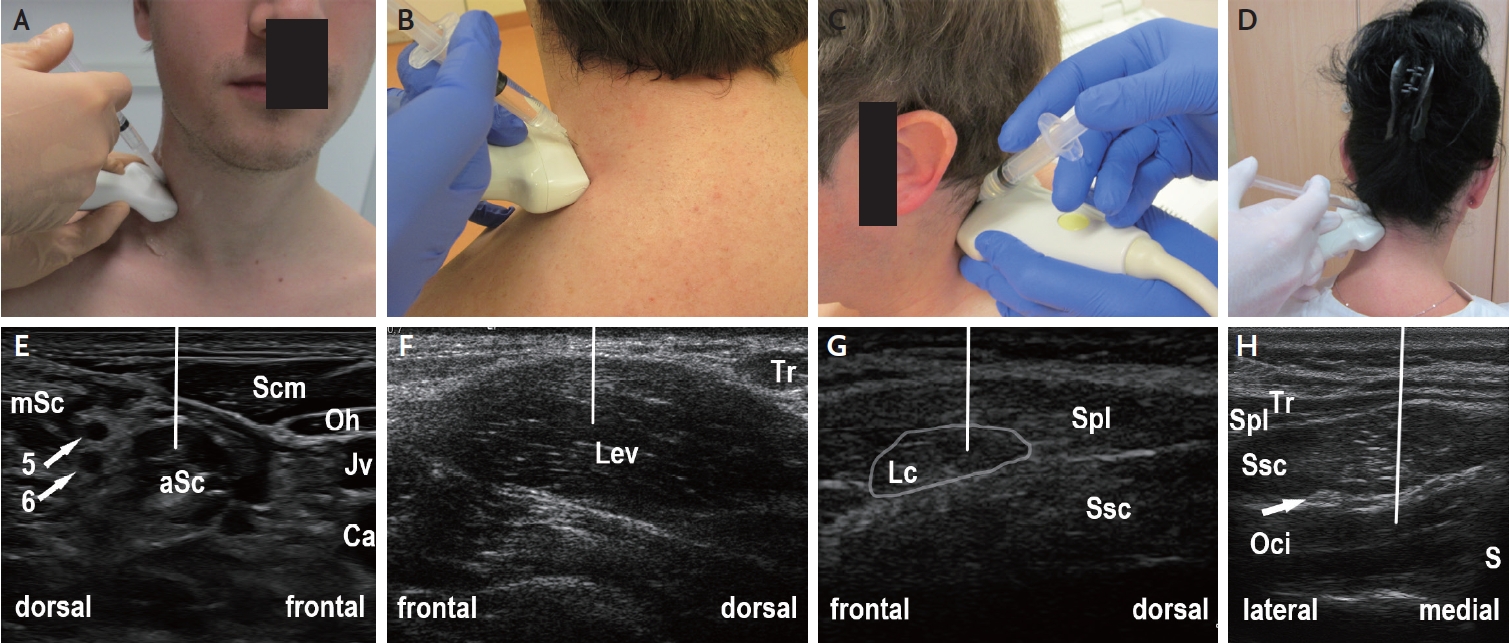
Especially for small and/or deeply located neck muscles (levator scapulae, splenius capitis, semispinalis capitis, longissimus capitis, obliquus capitis inferior, scalenus anterior, scalenus medius, longus colli) MUS-guided injection is recommendable (Fig. 4).6,71-73,75,76 A number of cohort studies demonstrated the safety and efficacy of MUS-guided BoNT injection in cervical dystonia.6,61,75-77 Recently the results of the first prospective, partially-blinded study comparing MUS-guided versus non-guided BoNT injection in cervical dystonia proved a clear additional benefit of MUS guidance.78 This may potentially allow for the decrease of individually applied BoNT doses, thereby reducing a potential risk of secondary immunogenic treatment failure which seems to be higher with injection of neck muscles as compared to muscles in other body regions.79

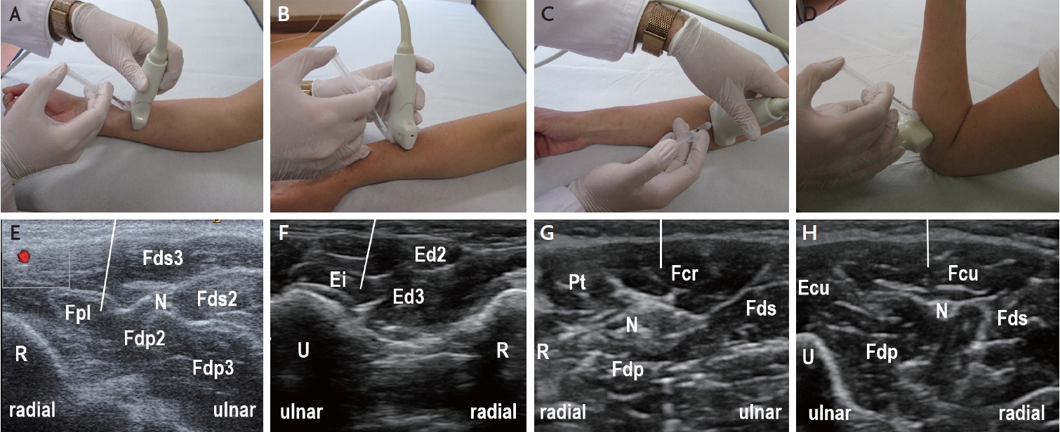
3. Task-related hand dystonia

Task-related dystonia (writer’s cramp, musician’s dystonia) involves forearm muscles that are dystonic only during specific activities. Therefore the therapeutic window of BoNT is small, and unintended paresis may occur. While there are no controlled studies assessing the use of MUS-guidance, there is expert consensus that a guidance technique (MUS or electrical stimulation) should be applied.6,80,81 A recent study found similar benefits with both guidance techniques, however favorable comfort of patients treated with MUS-guidance (Fig. 5).82 In many of our patients with writer’s cramp the BoNT dose could be reduced when switching from manual application to MUS-guidance.6

4. Cerebral palsy

Children and adolescents with cerebral palsy were among the first populations in whom MUS-guided injection techniques have been systematically applied.83 According to a European consensus statement,84 confirmed by subsequent systematic review,85 children with cerebral palsy should generally receive BoNT injections using EMG-guidance or MUS-guidance. Because of better tolerability, MUS-guidance is preferable in children. This applies also to adolescents and adults with cerebral palsy since chronic spasticity often led to muscular atrophy and joint deformation, requiring imaging-guided injection. MUS-guided BoNT injection provides a clear functional benefit to the patients.86 Findings of a recent sono-elastography study suggest that the treatment effect can be prolonged with extracorporeal shock wave therapy immediately following the BoNT injection.87

5. Poststroke spasticity

The use of intramuscular BoNT injection in poststroke spasticity is well established.88 Several small prospective randomised studies demonstrated superiority of MUS-guidance over the non-guided injection technique in patients with upper limb or lower limb spasticity.89-91 These findings were corroborated by observational studies.92-95 However, in another single-blinded, randomized controlled study the superiority of MUS-guidance could not be confirmed.96 In our experience MUS-guidance of BoNT injection in distinct muscles (Fig. 5, Table 4) may be favourable in patients with focal, or mild spasticity in whom precise targeting of a single muscle is required, and/or a functional benefit can be achieved.

6. Neurogenic thoracic outlet syndrome

Neurogenic thoracic outlet syndrome (nTOS) is a rare condition caused by compression of the brachial plexus at the thoracic outlet and presents with arm pain, paresthesias, and sometimes paresis of the hand. A minority of patients require decompressive surgery, however in many cases conservative treatment can be sufficient.97 Numerous case and cohort studies have shown a benefit of BoNT injection in the anterior scalene (Fig. 4A, 4E) and/or the pectoralis minor muscle.97 On the other hand, a double-blind, randomized, controlled trial did not find a significant improvement in pain or symptom reduction in nTOS patients.98 This study has been criticized because of EMG-guidance rather than imaging guidance, the very long symptom duration in the patients, and the inclusion of patients with mild symptoms at baseline.97 The results of a more recent study support the use of MUS-guided BoNT-injection.99

7. Piriformis syndrome

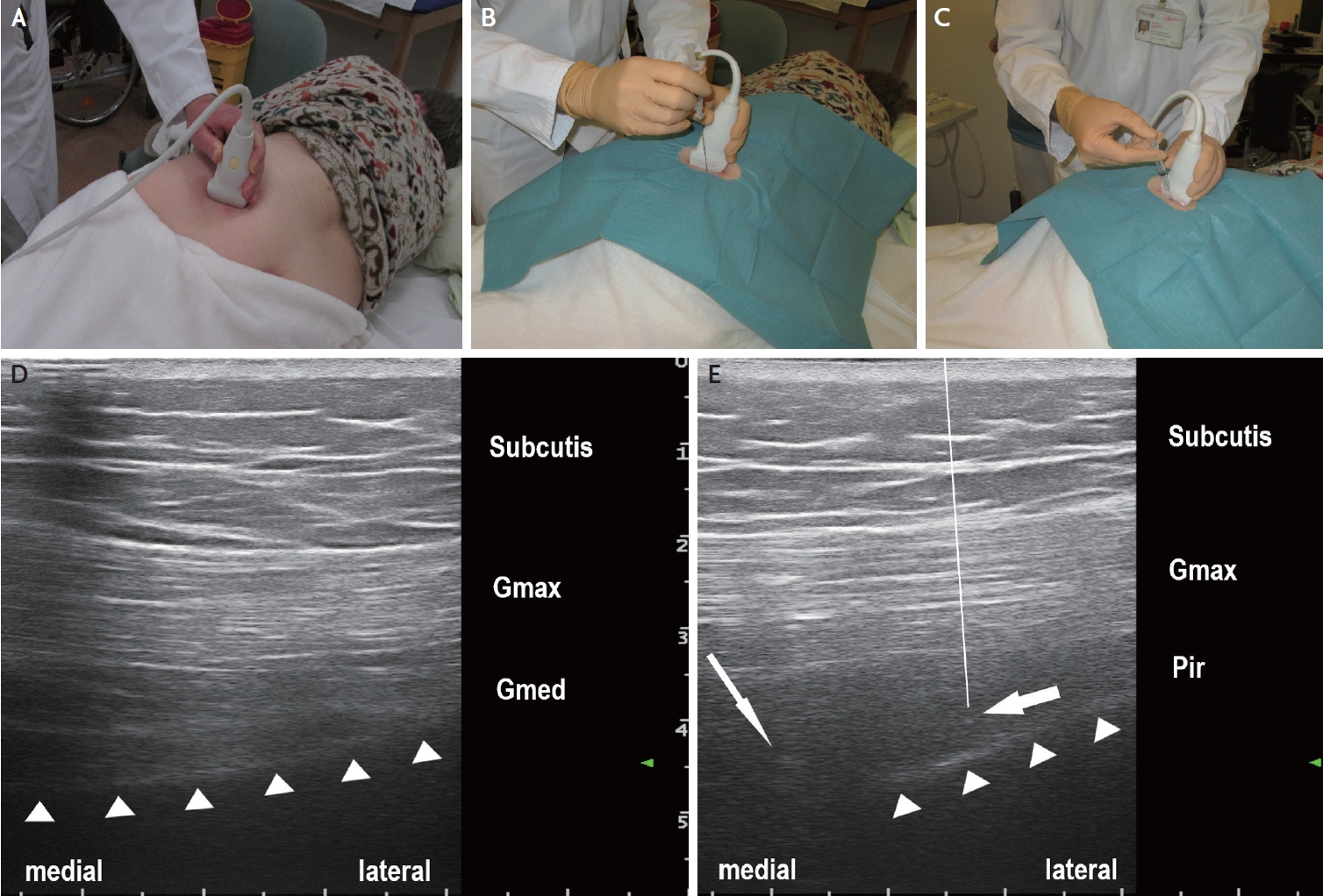
Piriformis syndrome is a neuromuscular condition caused by the entrapment of the sciatic nerve at the level of the piriformis muscle. It is diagnosed clinically by use of a set of specific tests, including the increase of pain during the FAIR (flexion, adduction, internal rotation) maneuver.100 This syndrome can well be treated with BoNT injection in the piriformis muscle. MUS-guided BoNT injection is more effective compared to MUS-guided injection of lidocaine or local ozone.100 Partial weakening of the piriformis muscle caused by the neurotoxin leads to decompression of the sciatic nerve which usually results in complete relief from pain. Usually this injection is done under computed tomography (CT-) guidance, which however necessitates the patients exposure to radiation and discomfort due to their prolonged positioning on the CT table. Moreover the sciatic nerve cannot always be visualized on CT. For MUS-guidance usually a 2–5 MHz curvilinear array transducer is used,100,101 however, in slender patients also a 6–13(–18) MHz linear array transducer is feasible (Fig. 6).100,102 This application requires higher intensity of antiseptic measures, the use of a long injection needle (e.g., a 120-mm, 25-gauge spinal needle), and should be performed by a physician who is well-experienced in conducting MUS-guided procedures.

CONCLUSION

MUS is increasingly used in neurology, neuropediatrics, neurosurgery and neuroradiology for the diagnosis of neuromuscular disorders. Especially, MUS is superior to other diagnostic tools in detecting fasciculations in motor neuron diseases and in revealing ICUAW. In addition MUS is an elegant tool for guiding needle biopsy and targeting intramuscular BoNT injection. Modern ultrasound technologies such as sono-elastography and high-definition ultrasound microvasculature imaging may allow for novel diagnostic and therapeutic uses.

NOTES

Ethics Statement
The Institutional Review Board process and patient consents were not proceeded because of a review article.
Availability of Data and Material
The data supporting the findings of this review are available from the author upon request.
Sources of Funding
None.
Conflicts of Interest
The author has received speaker honoraria and travel reimbursement from Ipsen Pharma, Merz Pharma, and Allergan/Abbvie, and a research grant from Merz Pharma. He serves as Joint Editor-in-Chief of Ultraschall in der Medizin/European Journal of Ultrasound.

Acknowledgments

None.

Fig. 1.
Muscle ultrasound findings in different neuromuscular disorders. (A) Normal forearm muscles with low, homogenous echogenicity. (B) Patient with amyotrophic lateral sclerosis. Note slightly increased echogenicity of Br, and markedly increased echogenicity of Fdp, with small areas of normal tissue inside (moth-eaten pattern, arrow). (C) Patient with facio-scapulo-humeral muscular dystrophy. Note homogenous, markedly increased echogenicity of the Bc. (D) patient with myositis. Note the patchy increase of echogenicity (arrows) in the Bb and Bc. Bb, biceps brachii; Bc, brachialis; Br, brachioradialis; Ecr, extensor carpi radialis; Fcr, flexor carpi radialis; Fdp, flexor digitorum profundus; H, humerus; Pt, pronator teres; R, radius; Sup, supinator.
jnn-2023-00133f1.jpg
Fig. 2.
Qualitative (visual) grading of sonographic muscle changes on the Heckmatt scale. (A, E) Normal echo-intensity of the muscle with normal echo-intensity and full visualization of the adjacent bone cortex (grade 1). (B, F) Moderately increased echo-intensity of the muscle with normal echo-intensity and full visualization of the adjacent bone cortex (grade 2). (C, G) Markedly increased echo-intensity of the muscle with altered echo-signal and incomplete visualization of the adjacent bone cortex (grade 3). (D, H) Strongly increased echo-intensity of the muscle with complete loss of bone cortex echo-signal (grade 4). Bc, brachialis; Br, brachioradialis; H, humerus; T, tibia; Ta, tibialis anterior.
jnn-2023-00133f2.jpg
Fig. 3.
Detection of fasciculations on muscle ultrasound in patients with motor neuron diseases. (A) Positioning of the ultrasound transducer for visualization of tongue muscles displayed in (B). (B) Coronal B-mode sonogram (right panel) and corresponding M-mode sonogram (left panel) through tongue muscles. The rhombi denote fasciculations in the Gg. (C) Axial B-mode sonogram (right panel) and corresponding M-mode sonogram (left panel) through lumbar paraspinal muscles. The rhombi denote fasciculations in the Es. (D) Positioning of the ultrasound transducer for visualization of radial forearm muscles displayed in (E). (E) Axial B-mode sonogram (right panel) and corresponding M-mode sonogram (left panel) through proximal forearm muscles. The rhombi denote fasciculations in the Fdp and Br. (F) Axial B-mode sonogram (right panel) and corresponding M-mode sonogram (left panel) through anterior thigh muscles. The rhombi denote fasciculations in the Rf. Br, brachioradialis; Es, erector spinae; Gg, genioglossus; F, femur; Fdp, flexor digitorum profundus; Hg, hyoglossus; Mh, mylohyoid; Pt, pronator teres; Rf, rectus femoris; S, spinous process; Vi, vastus intermedius.
jnn-2023-00133f3.jpg
Fig. 4.
Muscle ultrasound of the lateral and dorsal neck region for guiding botulinum neurotoxin injection (off-plane injection technique). (A) Position of ultrasound transducer and syringe for guiding injection into the aSc. (B) Position of ultrasound transducer and syringe for guiding injection into the Lev. (C) Position of ultrasound transducer and syringe for guiding injection into the Lc. (D) Position of ultrasound transducer and syringe for guiding injection into the Oci. (E) Ultrasound image corresponding to (A) showing the lateral neck region. The aSc and mSc are discerned by visualizing the nerve roots of the brachial plexus (arrows, 5=5th cervical nerve root; 6=6th cervical nerve root). The line indicates the expected path of injection needle. (F) Ultrasound image corresponding to (B) showing the lateral neck region, with transducer position optimized for Lev injection. (G) Ultrasound image corresponding to (C) showing the lateral neck region, with transducer position optimized for Lc injection. (H) Ultrasound image corresponding to (D) showing the dorsal neck region, with transducer position optimized for Oci injection (arrow: greater occipital nerve). Note that the needle should be guided from medio-dorsal to slightly antero-ventral direction (indicated by the line) in order to avoid touching the vertebral artery. aSc, anterior scalene; Ca, common carotid artery; Jv, internal jugular vein; Lc, longissimus capitis; Lev, levator scapulae; mSc, middle scalene; Oci, obliquus capitis inferior; Oh, omohyoid; S, spinous process; Scm, sternocleidomastoid; Spl, splenius capitis; Ssc, semispinalis capitis; Tr, trapezius.
jnn-2023-00133f4.jpg
Fig. 5.
Muscle ultrasound of the forearm muscles for guiding botulinum neurotoxin injection (off-plane injection technique). (A) Position of ultrasound transducer and syringe for guiding injection into the Fpl. (B) Position of ultrasound transducer and syringe for guiding injection into the Ei. (C) Position of ultrasound transducer and syringe for guiding injection into the Fcr. (D) Position of ultrasound transducer and syringe for guiding injection into the Fcu. (E) Ultrasound image corresponding to (A) showing the distal forearm flexors. For guiding the injection into the Fpl, the radial artery (red) and the median nerve (N) should be clearly displayed. The line indicates the expected path of injection needle. (F) Ultrasound image corresponding to (B) showing the distal forearm extensors, with transducer position optimized for Ei injection. (G) Ultrasound image corresponding to (C) showing the proximal forearm flexors, with transducer position optimized for Fcr injection (N: median nerve). (H) Ultrasound image corresponding to (D) showing the proximal forearm flexors, with transducer position optimized for Fcu injection (N: ulnar nerve). Ecu, extensor carpi ulnaris; Ei, extensor indicis; Ed, extensor digitorum; Fcr, flexor carpi radialis; Fcu, flexor carpi ulnaris; Fds, flexor digitorum superficialis; Fdp, flexor digitorum profundus; Fpl, flexor pollicis longus; N, nerve; Pt, pronator teres; R, radius; U, ulna.
jnn-2023-00133f5.jpg
Fig. 6.
Ultrasound-guided botulinum neurotoxin injection into the piriformis muscle in a patient with severe, disabling piriformis syndrome. (A) Ultrasound transducer positioning for identification of the piriformis muscle. (B) Ultrasound-guided insertion of injection needle (here: 22-gauge/0.70×88 mm spinal needle) performed under antiseptic conditions. (C) Ultrasound-guided injection. (D) Semi-axial ultrasound imaging plane showing the gluteus maximus (Gmax) and the gluteus medius (Gmed) muscles and the iliac bone (triangles) cranial to the greater sciatic notch. (E) Semi-axial ultrasound imaging plane slightly caudal to the plane presented in (D). The piriformis muscle (Pir) is located with its lateral part between the gluteus maximus muscle (Gmax) and the iliac bone (triangles), and with its medial part between the gluteus maximus muscle and the greater sciatic notch (thin arrow: sciatic nerve). The thick arrow indicates the position of the needle tip in the piriformis muscle. The line indicates the path of needle which is not completely visible on the ultrasound image since an off-plane injection technique is used (with only the needle tip visualized in the target region).
jnn-2023-00133f6.jpg
Table 1.
Comparison of ultrasound frequency, image resolution, and imaging depth
Frequency (MHz) Image resolution* axial (mm)/lateral (mm) Imaging depth* (mm) Exemplary muscles
5 0.30/1.0 120 Hamstring muscles
10 0.16/0.6 60 Calf muscles
15 0.12/0.4 30 Forearm muscles
20 0.07/0.2 15 Mimic muscles
30 0.05/0.18 10 Thenar muscles

* Given are rough values that can vary depending on tissue composition, quality of ultrasound transducer, and image postprocessing technology (modified from Walter12).

Table 2.
Muscle ultrasound findings in neuromuscular disorders
Entity EI within an affected muscle Pattern of involved muscles
MND/Neuropathy Moderately increased (denervated muscle) All muscles which are innervated by the degenerated motor neurons / nerves
Moth-eaten pattern (normal EI of re-innervated muscle foci)
Dermato-/Polymyositis/Necrotizing auto-immune myopathy Moderately increased (whole muscle or focally/patchy) All muscles in the affected body region
Inclusion body myositis Markedly increased (homogenous increase) Bilateral deep forearm flexors (esp. FDP), quadriceps femoris and gastrocnemius (often asymmetric)
Muscular dystrophy Markedly increased (homogenous with “ground glass” appearance, sometimes deeper areas of normal EI due to increased attenuation) In early disease stages selected muscles, in late disease stages more generalized
Congenital myopathy Normal to markedly increased (homogenous increase) Depending on entity (focal or generalized)
Metabolic myopathy Normal to slightly increased (homogenous increase) Often generalized

Modified from Pillen et al.22 and Mah and van Alfen.23

EI denotes echo-intensity; FDP, flexor digitorum profundus; MND, motor neuron disease.

Table 3.
The Heckmatt scale for visual grading of muscle echo-intensity
Grade Finding
Grade 1 Normal echo-intensity of the muscle with normal echo-intensity and full visualization of the adjacent bone cortex
Grade 2 Moderately increased echo-intensity of the muscle and distinct bone echo with normal echo-intensity and full visual- ization of the adjacent bone cortex
Grade 3 Markedly increased echo-intensity of the muscle and reduced bone echo with altered echo-signal and incomplete visualization of the adjacent bone cortex
Grade 4 Strongly increased echo-intensity of the muscle with complete loss of bone cortex echo-signal.

Scale proposed by Heckmatt et al.25

Table 4.
Muscles most frequently injected with MUS guidance in the author’s clinic
Muscle Diseases BoNT-A dose (MU)*
Head/neck
Lateral pterygoid Bruxism, oromandibular dystonia 25 (8–60)
Digastric Oromandibular dystonia 15 (10–20)
Anterior scalene nTOS; cervical dystonia 20 (12–50); 35 (20–80)
Longissimus capitis Cervical dystonia 30 (10–60)
Splenius capitis Cervical dystonia 55 (10–200)
Obliquus capitis inferior Cervical dystonia 30 (10–60)
Shoulder/arm
Pectoralis minor nTOS 20 (12–50)
Pronator teres Poststroke spasticity 30 (10–100)
Flexor carpi radialis Poststroke spasticity; hand dystonia 50 (30–100); 30 (5–60)
Flexor carpi ulnaris Poststroke spasticity; hand dystonia 50 (30–100); 30 (5–60)
Single superfic. finger flexors Poststroke spasticity; hand dystonia 25 (10–50); 15 (5–40)
Single deep finger flexors Poststroke spasticity; hand dystonia 25 (10–50); 15 (5–40)
Single finger extensors Hand dystonia 5 (2.5–20)
Extensor indicis Hand dystonia 8 (5–10)
Pronator quadratus Hand dystonia 15 (5–40)
Pelvis/Leg
Piriformis Piriformis syndrome 100 (50–150)
Iliopsoas Cerebral palsy 50 (25–200)
Rectus femoris Cerebral palsy 60 (20–150)
Adductor longus Cerebral palsy 50 (20–120)
Semimembranosus Cerebral palsy 50 (20–140)
Tibialis posterior Poststroke spasticity, cerebral palsy 70 (20–140)
Gastrocnemius, caput med. Poststroke spasticity, cerebral palsy 70 (20–220)
Gastrocnemius, caput laterale Poststroke spasticity, cerebral palsy 40 (20–80)
Soleus Poststroke spasticity, cerebral palsy 40 (20–100)
Extensor hallucis longus Poststroke spasticity 40 (20–100)
Flexor hallucis longus Poststroke spasticity 40 (10–100)
Flexor digitorum longus Poststroke spasticity 50 (20–80)

Values are presented as typical dose (dose limits).

BoNT-A refers to botulinum neurotoxin type A; MU, mouse units; nTOS, neurogenic thoracic outlet syndrome.

* The BoNT-A doses (typical dose, dose limits; modified from Jost63 and Dressler et al.64 given per muscle per treatment session refer to the preparations onabotulinumtoxinA (Botox®; Allergan/Abbvie, Irvine, CA, USA) and incobotulinumtoxinA (Xeomin®; Merz Pharmaceuticals, Frankfurt/M, Germany). Other BoNT-A preparations such as abobotulinumtoxinA (Dysport®; Ipsen, Billancourt, France) and prabotulinumtoxinA (Nabota®; Daewoong Pharmaceutical, Seoul, Korea) may be used, however, dose conversion ratios apply due to their different potency labelling. Note that for the different BoNT-A preparations part of the applications listed here are off-label, depending on the marketing authorization by the responsible medicines agency.

Usually task-related dystonia (writer’s cramp, musician dystonia).

Usually task-related dystonia, or post-traumatic hand dystonia (causalgia).

REFERENCES

1. Heckmatt JZ, Dubowitz V, Leeman S. Detection of pathological change in dystrophic muscle with B-scan ultrasound imaging. Lancet. 1980;1:1389-1390.
crossref pmid
2. Fischer AQ, Carpenter DW, Hartlage PL, Carroll JE, Stephens S. Muscle imaging in neuromuscular disease using computerized real-time sonography. Muscle Nerve. 1988;11:270-275.
crossref pmid
3. Reimers CD, Fleckenstein JL, Witt TN, Müller-Felber W, Pongratz DE. Muscular ultrasound in idiopathic inflammatory myopathies of adults. J Neurol Sci. 1993;116:82-92.
crossref pmid
4. Reimers CD, Schlotter B, Eicke BM, Witt TN. Calf enlargement in neuromuscular diseases: a quantitative ultrasound study in 350 patients and review of the literature. J Neurol Sci. 1996;143:46-56.
crossref pmid
5. Walker FO, Cartwright MS, Wiesler ER, Caress J. Ultrasound of nerve and muscle. Clin Neurophysiol. 2004;115:495-507.
crossref pmid
6. Walter U, Dressler D. Ultrasound-guided botulinum toxin injections in neurology: technique, indications and future perspectives. Expert Rev Neurother. 2014;14:923-936.
crossref pmid
7. Mah JK, van Alfen N. Neuromuscular ultrasound: Clinical applications and diagnostic values. Can J Neurol Sci. 2018;45:605-619.
crossref pmid
8. Hannaford A, Vucic S, van Alfen N, Simon NG. Muscle ultrasound in hereditary muscle disease. Neuromuscul Disord. 2022;32:851-863.
crossref pmid
9. Tawfik EA, Cartwright MS, Grimm A, Boon AJ, Kerasnoudis A, Preston DC, et al. Guidelines for neuromuscular ultrasound training. Muscle Nerve. 2019;60:361-366.
crossref pmid pdf
10. Peetrons P. Ultrasound of muscles. Eur Radiol. 2002;12:35-43.
crossref pmid pdf
11. Volk GF, Sauer M, Pohlmann M, Guntinas-Lichius O. Reference values for dynamic facial muscle ultrasonography in adults. Muscle Nerve. 2014;50:348-357.
crossref pmid
12. Walter U. How small can small nerves be for diagnostic ultrasonography? Ultraschall Med. 2019;40:400-402.
crossref pmid
13. Schmidt WA, Backhaus M, Sattler H, Kellner H. [Imaging techniques in rheumatology: sonography in rheumatoid arthritis]. Z Rheumatol. 2003;62:23-33 German.
pmid
14. Boon AJ, Oney-Marlow TM, Murthy NS, Harper CM, McNamara TR, Smith J. Accuracy of electromyography needle placement in cadavers: non-guided vs. ultrasound guided. Muscle Nerve. 2011;44:45-49.
crossref pmid
15. Mayans D, Cartwright MS, Walker FO. Neuromuscular ultrasonography: quantifying muscle and nerve measurements. Phys Med Rehabil Clin N Am. 2012;23:133-148.
crossref pmid pmc
16. To EW, Ahuja AT, Ho WS, King WW, Wong WK, Pang PC, et al. A prospective study of the effect of botulinum toxin A on masseteric muscle hypertrophy with ultrasonographic and electromyographic measurement. Br J Plast Surg. 2001;54:197-200.
crossref pmid
17. Stokes M, Hides J, Elliott J, Kiesel K, Hodges P. Rehabilitative ultrasound imaging of the posterior paraspinal muscles. J Orthop Sports Phys Ther. 2007;37:581-595.
crossref pmid
18. English C, Fisher L, Thoirs K. Reliability of real-time ultrasound for measuring skeletal muscle size in human limbs in vivo: a systematic review. Clin Rehabil. 2012;26:934-944.
crossref pmid pdf
19. Alfen NV, Gilhuis HJ, Keijzers JP, Pillen S, Van Dijk JP. Quantitative facial muscle ultrasound: feasibility and reproducibility. Muscle Nerve. 2013;48:375-380.
crossref pmid pdf
20. Qin X, Fei B. Measuring myofiber orientations from high-frequency ultrasound images using multiscale decompositions. Phys Med Biol. 2014;59:3907-3924.
crossref pmid pmc pdf
21. Pillen S, Tak RO, Zwarts MJ, Lammens MM, Verrijp KN, Arts IM, et al. Skeletal muscle ultrasound: correlation between fibrous tissue and echo intensity. Ultrasound Med Biol. 2009;35:443-446.
crossref pmid
22. Pillen S, Arts IM, Zwarts MJ. Muscle ultrasound in neuromuscular disorders. Muscle Nerve. 2008;37:679-693.
crossref pmid
23. Mah JK, van Alfen N. Neuromuscular ultrasound: Clinical applications and diagnostic values. Can J Neurol Sci. 2018;45:605-619.
crossref pmid
24. Paramalingam S, Needham M, Harris S, O’Hanlon S, Mastaglia F, Keen H. Muscle B mode ultrasound and shearwave elastography in idiopathic inflammatory myopathies (SWIM): criterion validation against MRI and muscle biopsy findings in an incident patient cohort. BMC Rheumatol. 2022;6:47.
crossref pmid pmc pdf
25. Heckmatt JZ, Leeman S, Dubowitz V. Ultrasound imaging in the diagnosis of muscle disease. J Pediatr. 1982;101:656-660.
crossref pmid
26. Zaidman CM, Wu JS, Kapur K, Pasternak A, Madabusi L, Yim S, et al. Quantitative muscle ultrasound detects disease progression in Duchenne muscular dystrophy. Ann Neurol. 2017;81:633-640.
crossref pmid pmc pdf
27. Goselink RJM, Schreuder THA, Mul K, Voermans NC, Erasmus CE, van Engelen BGM, et al. Muscle ultrasound is a responsive biomarker in facioscapulohumeral dystrophy. Neurology. 2020;94:e1488-e1494.
crossref pmid
28. Vill K, Sehri M, Müller C, Hannibal I, Huf V, Idriess M, et al. Qualitative and quantitative muscle ultrasound in patients with Duchenne muscular dystrophy: Where do sonographic changes begin? Eur J Paediatr Neurol. 2020;28:142-150.
crossref pmid
29. Abdelnaby R, Mohamed KA, Elgenidy A, Sonbol YT, Bedewy MM, Aboutaleb AM, et al. Muscle sonography in inclusion body myositis: A systematic review and meta-analysis of 944 measurements. Cells. 2022;11:600.
crossref pmid pmc
30. Li M, Guo R, Tang X, Huang S, Qiu L. Quantitative assessment of muscle properties in polymyositis and dermatomyositis using high-frequency ultrasound and shear wave elastography. Quant Imaging Med Surg. 2023;13:428-440.
crossref pmid pmc
31. Hermans G, Van den Berghe G. Clinical review: intensive care unit acquired weakness. Crit Care. 2015;19:274.
crossref pmid pmc pdf
32. Senger D, Erbguth F. [Critical illness myopathy and polyneuropathy]. Med Klin Intensivmed Notfmed. 2017;112:589-596 German.
crossref pmid pmc pdf
33. Connolly BA, Jones GD, Curtis AA, Murphy PB, Douiri A, Hopkinson NS, et al. Clinical predictive value of manual muscle strength testing during critical illness: an observational cohort study. Crit Care. 2013;17:R229.
crossref pmid pmc
34. Grimm A, Teschner U, Porzelius C, Ludewig K, Zielske J, Witte OW, et al. Muscle ultrasound for early assessment of critical illness neuromyopathy in severe sepsis. Crit Care. 2013;17:R227.
crossref pmid pmc
35. Kelmenson DA, Quan D, Moss M. What is the diagnostic accuracy of single nerve conduction studies and muscle ultrasound to identify critical illness polyneuromyopathy: a prospective cohort study. Crit Care. 2018;22:342.
crossref pmid pmc pdf
36. Hadda V, Kumar R, Khilnani GC, Kalaivani M, Madan K, Tiwari P, et al. Trends of loss of peripheral muscle thickness on ultrasonography and its relationship with outcomes among patients with sepsis. J Intensive Care. 2018;6:81.
crossref pmid pmc pdf
37. Patejdl R, Walter U, Rosener S, Sauer M, Reuter DA, Ehler J. Muscular ultrasound, syndecan-1 and procalcitonin serum levels to assess intensive care unit-acquired weakness. Can J Neurol Sci. 2019;46:234-242.
crossref pmid
38. Naoi T, Morita M, Koyama K, Katayama S, Tonai K, Sekine T, et al. Upper arm muscular echogenicity predicts intensive care unit-acquired weakness in critically Ill patients. Prog Rehabil Med. 2022;7:20220034.
crossref pmid pmc
39. Klawitter F, Walter U, Patejdl R, Endler J, Reuter DA, Ehler J. Sonographic evaluation of muscle echogenicity for the detection of intensive care unit-acquired weakness: A pilot single-center prospective cohort study. Diagnostics (Basel). 2022;12:1378.
crossref pmid pmc
40. Paolo F, Valentina G, Silvia C, Tommaso P, Elena C, Martin D, et al. The possible predictive value of muscle ultrasound in the diagnosis of ICUAW in long-term critically ill patients. J Crit Care. 2022;71:154104.
crossref pmid
41. Zhang W, Wu J, Gu Q, Gu Y, Zhao Y, Ge X, et al. Changes in muscle ultrasound for the diagnosis of intensive care unit acquired weakness in critically ill patients. Sci Rep. 2021;11:18280.
crossref pmid pmc pdf
42. Witteveen E, Sommers J, Wieske L, Doorduin J, van Alfen N, Schultz MJ, et al. Diagnostic accuracy of quantitative neuromuscular ultrasound for the diagnosis of intensive care unit-acquired weakness: a cross-sectional observational study. Ann Intensive Care. 2017;7:40.
crossref pmid pmc pdf
43. Puthucheary ZA, Phadke R, Rawal J, McPhail MJ, Sidhu PS, Rowlerson A, et al. Qualitative ultrasound in acute critical illness muscle wasting. Crit Care Med. 2015;43:1603-1611.
crossref pmid
44. Hernández-Socorro CR, Saavedra P, López-Fernández JC, Lübbe-Vazquez F, Ruiz-Santana S. Novel high-quality sonographic methods to diagnose muscle wasting in longstay critically Ill patients: Shear wave elastography, superb microvascular imaging and contrast-enhanced ultrasound. Nutrients. 2021;13:2224.
crossref pmid pmc
45. Abraham A, Drory VE, Fainmesser Y, Algom AA, Lovblom LE, Bril V. Muscle thickness measured by ultrasound is reduced in neuromuscular disorders and correlates with clinical and electrophysiological findings. Muscle Nerve. 2019;60:687-692.
crossref pmid pdf
46. Arts IM, Schelhaas HJ, Verrijp KC, Zwarts MJ, Overeem S, van der Laak JA, et al. Intramuscular fibrous tissue determines muscle echo intensity in amyotrophic lateral sclerosis. Muscle Nerve. 2012;45:449-450.
crossref pmid
47. Buonsenso D, Berti B, Palermo C, Leone D, Ferrantini G, De Sanctis R, et al. Ultrasound assessment of diaphragmatic function in type 1 spinal muscular atrophy. Pediatr Pulmonol. 2020;55:1781-1788.
crossref pmid pdf
48. Hobson-Webb LD, Simmons Z. Ultrasound in the diagnosis and monitoring of amyotrophic lateral sclerosis: A review. Muscle Nerve. 2019;60:114-123.
crossref pmid pdf
49. Hermann W, Langner S, Freigang M, Fischer S, Storch A, Günther R, et al. Affection of respiratory muscles in ALS and SMA. J Clin Med. 2022;11:1163.
crossref pmid pmc
50. Seok HY, Park J, Kim YH, Oh KW, Kim SH, Kim BJ. Split hand muscle echo intensity index as a reliable imaging marker for differential diagnosis of amyotrophic lateral sclerosis. J Neurol Neurosurg Psychiatry. 2018;89:943-948.
crossref pmid
51. Dos Santos MAR, Brighente SF, Massignan A, Tenório RB, Makariewicz LL, Moreira AL, et al. Accuracy of muscle fasciculations for the diagnosis of later-onset spinal muscle atrophy. Neuromuscul Disord. 2022;32:763-768.
crossref pmid
52. Grimm A, Prell T, Décard BF, Schumacher U, Witte OW, Axer H, et al. Muscle ultrasonography as an additional diagnostic tool for the diagnosis of amyotrophic lateral sclerosis. Clin Neurophysiol. 2015;126:820-827.
crossref pmid
53. Takamatsu N, Nodera H, Mori A, Maruyama-Saladini K, Osaki Y, Shimatani Y, et al. Which muscle shows fasciculations by ultrasound in patients with ALS? J Med Invest. 2016;63:49-53.
crossref pmid
54. Noto YI, Shibuya K, Shahrizaila N, Huynh W, Matamala JM, Dharmadasa T, et al. Detection of fasciculations in amyotrophic lateral sclerosis: The optimal ultrasound scan time. Muscle Nerve. 2017;56:1068-1071.
crossref pmid pdf
55. Ma J, Wen Q, Pang X, Huang S, Zhang J, Wang J, et al. Fasciculation score: a sensitive biomarker in amyotrophic lateral sclerosis. Neurol Sci. 2021;42:4657-4666.
crossref pmid pdf
56. Hagiwara Y, Shimizu T, Yanagisawa T, Akasu Y, Kaburagi K, Kikuchi T, et al. Utility of transoral motion-mode ultrasonography to detect tongue fasciculation in patients with amyotrophic lateral sclerosis. Muscle Nerve. 2021;63:909-913.
crossref pmid pdf
57. Ludolph A, Drory V, Hardiman O, Nakano I, Ravits J, Robberecht W, et al. A revision of the El Escorial criteria - 2015. Amyotroph Lateral Scler Frontotemporal Degener. 2015;16:291-292.
crossref pmid
58. Lagnau P, Lo A, Sandarage R, Alter K, Picelli A, Wissel J, et al. Ergonomic recommendations in ultrasound-guided botulinum neurotoxin chemodenervation for spasticity: An international expert group opinion. Toxins (Basel). 2021;13:249.
crossref pmid pmc
59. Schramm A, Bäumer T, Fietzek U, Heitmann S, Walter U, Jost WH. Relevance of sonography for botulinum toxin treatment of cervical dystonia: an expert statement. J Neural Transm (Vienna). 2015;122:1457-1463.
crossref pmid pmc pdf
60. Schramm A, Huber D, Möbius C, Münchau A, Kohl Z, Bäumer T. Involvement of obliquus capitis inferior muscle in dystonic head tremor. Parkinsonism Relat Disord. 2017;44:119-123.
crossref pmid
61. Fietzek UM, Nene D, Schramm A, Appel-Cresswell S, Košutzká Z, Walter U, et al. The role of ultrasound for the personalized botulinum toxin treatment of cervical dystonia. Toxins (Basel). 2021;13:365.
crossref pmid pmc
62. Spina S, Facciorusso S, Botticelli C, Intiso D, Ranieri M, Colamaria A, et al. Ultrasonographic evaluation of three approaches for botulinum toxin injection into tibialis posterior muscle in chronic stroke patients with equinovarus foot: An observational study. Toxins (Basel). 2021;13:829.
crossref pmid pmc
63. Jost W. Atlas of Botulinum Toxin Injection. 3rd ed. Berlin: Quintessence Publishing; 2019.

64. Dressler D, Altavista MC, Altenmueller E, Bhidayasiri R, Bohlega S, Chana P, et al. Consensus guidelines for botulinum toxin therapy: general algorithms and dosing tables for dystonia and spasticity. J Neural Transm (Vienna). 2021;128:321-335.
crossref pmid pmc pdf
65. Kong KH, Shuen-Loong T, Tay MRJ, Lui WL, Rajeswaran DK, Kim J. Ultrasound assessment of changes in muscle architecture of the brachialis muscle after stroke-a prospective study. Arch Rehabil Res Clin Transl. 2022;4:100215.
crossref pmid pmc
66. Kesikburun S, Yaşar E, Adıgüzel E, Güzelküçük Ü, Alaca R, Tan AK. Assessment of spasticity with sonoelastography following stroke: A feasibility study. PM R. 2015;7:1254-1260.
crossref pmid pdf
67. Gao J, Rubin JM, Chen J, O’Dell M. Ultrasound elastography to assess botulinum toxin a treatment for post-stroke spasticity: A feasibility study. Ultrasound Med Biol. 2019;45:1094-1102.
crossref pmid
68. Song Y, Zhang TJ, Li Y, Gao Y. Application of real-time shear wave elastography in the assessment of torsional cervical dystonia. Quant Imaging Med Surg. 2019;9:662-670.
crossref pmid pmc
69. Henzel MK, Munin MC, Niyonkuru C, Skidmore ER, Weber DJ, Zafonte RD. Comparison of surface and ultrasound localization to identify forearm flexor muscles for botulinum toxin injections. PM R. 2010;2:642-646.
crossref pmid pmc pdf
70. Picelli A, Roncari L, Baldessarelli S, Berto G, Lobba D, Santamato A, et al. Accuracy of botulinum toxin type A injection into the forearm muscles of chronic stroke patients with spastic flexed wrist and clenched fist: manual needle placement evaluated using ultrasonography. J Rehabil Med. 2014;46:1042-1045.
crossref pmid
71. Ko YD, Yun SI, Ryoo D, Chung ME, Park J. Accuracy of ultrasound-guided and non-guided botulinum toxin injection into neck muscles involved in cervical dystonia: A cadaveric study. Ann Rehabil Med. 2020;44:370-377.
crossref pmid pmc pdf
72. Kreisler A, Gerrebout C, Defebvre L, Demondion X. Accuracy of non-guided versus ultrasound-guided injections in cervical muscles: a cadaver study. J Neurol. 2021;268:1894-1902.
crossref pmid pdf
73. Oh D, Lee HS. Atypical course of vertebral artery identified by ultrasound prescan before performing a stellate ganglion block. J Med Ultrasound. 2021;30:143-145.
crossref pmid pmc
74. Farrell M, Karp BI, Kassavetis P, Berrigan W, Yonter S, Ehrlich D, et al. Management of anterocapitis and anterocollis: A novel ultrasound guided approach combined with electromyography for botulinum toxin injection of longus colli and longus capitis. Toxins (Basel). 2020;12:626.
crossref pmid pmc
75. Lee IH, Yoon YC, Sung DH, Kwon JW, Jung JY. Initial experience with imaging-guided intramuscular botulinum toxin injection in patients with idiopathic cervical dystonia. AJR Am J Roentgenol. 2009;192:996-1001.
crossref pmid
76. Walter U, Dudesek A, Fietzek UM. A simplified ultrasonography-guided approach for neurotoxin injection into the obliquus capitis inferior muscle in spasmodic torticollis. J Neural Transm (Vienna). 2018;125:1037-1042.
crossref pmid pdf
77. Huang L, Chen HX, Ding XD, Xiao HQ, Wang W, Wang H. Efficacy analysis of ultrasound-guided local injection of botulinum toxin type A treatment with orthopedic joint brace in patients with cervical dystonia. Eur Rev Med Pharmacol Sci. 2015;19:1989-1993.
pmid
78. Tyślerowicz M, Dulski J, Gawryluk J, Sławek J. Does ultrasound guidance improve the effectiveness of neurotoxin injections in patients with cervical dystonia? (A prospective, partially-blinded, clinical study). Toxins (Basel). 2022;14:674.
crossref pmid pmc
79. Walter U, Mühlenhoff C, Benecke R, Dressler D, Mix E, Alt J, et al. Frequency and risk factors of antibody-induced secondary failure of botulinum neurotoxin therapy. Neurology. 2020;94:e2109-e2120.
crossref pmid
80. Lim EC, Quek AM, Seet RC. Accurate targeting of botulinum toxin injections: how to and why. Parkinsonism Relat Disord. 2011;17 Suppl 1:S34-39.
crossref pmid
81. Catania S. How do I evaluate and inject for Writer’s cramp? Mov Disord Clin Pract. 2018;5:663.
crossref pmid pmc pdf
82. Lungu C, Nmashie A, George MC, Karp BI, Alter K, Shin S, et al. Comparison of ultrasound and electrical stimulation guidance for onabotulinum toxin-A injections: A randomized crossover study. Mov Disord Clin Pract. 2022;9:1055-1061.
pmid pmc
83. Berweck S, Feldkamp A, Francke A, Nehles J, Schwerin A, Heinen F. Sonography-guided injection of botulinum toxin A in children with cerebral palsy. Neuropediatrics. 2002;33:221-223.
crossref pmid
84. Heinen F, Desloovere K, Schroeder AS, Berweck S, Borggraefe I, van Campenhout A, et al. The updated European Consensus 2009 on the use of Botulinum toxin for children with cerebral palsy. Eur J Paediatr Neurol. 2010;14:45-66.
crossref pmid
85. Grigoriu AI, Dinomais M, Rémy-Néris O, Brochard S. Impact of injection-guiding techniques on the effectiveness of botulinum toxin for the treatment of focal spasticity and dystonia: A systematic review. Arch Phys Med Rehabil. 2015;96:2067-2078.e1.
crossref pmid
86. Kim SK, Rha DW, Park ES. Botulinum toxin type A injections impact hamstring muscles and gait parameters in children with flexed knee gait. Toxins (Basel). 2020;12:145.
crossref pmid pmc
87. Kwon DR, Kwon DG. Botulinum toxin A injection combined with radial extracorporeal shock wave therapy in children with spastic cerebral palsy: Shear wave sonoelastographic findings in the medial gastrocnemius muscle, preliminary study. Children (Basel). 2021;8:1059.
crossref pmid pmc
88. Sun LC, Chen R, Fu C, Chen Y, Wu Q, Chen R, et al. Efficacy and safety of botulinum toxin type A for limb spasticity after stroke: A meta-analysis of randomized controlled trials. Biomed Res Int. 2019;2019:8329306.
crossref pmid pmc pdf
89. Picelli A, Tamburin S, Bonetti P, Fontana C, Barausse M, Dambruoso F, et al. Botulinum toxin type A injection into the gastrocnemius muscle for spastic equinus in adults with stroke: a randomized controlled trial comparing manual needle placement, electrical stimulation and ultrasonography-guided injection techniques. Am J Phys Med Rehabil. 2012;91:957-964.
pmid
90. Picelli A, Lobba D, Midiri A, Prandi P, Melotti C, Baldessarelli S, et al. Botulinum toxin injection into the forearm muscles for wrist and fingers spastic overactivity in adults with chronic stroke: a randomized controlled trial comparing three injection techniques. Clin Rehabil. 2014;28:232-242.
crossref pmid pdf
91. Santamato A, Micello MF, Panza F, Fortunato F, Baricich A, Cisari C, et al. Can botulinum toxin type A injection technique influence the clinical outcome of patients with poststroke upper limb spasticity? A randomized controlled trial comparing manual needle placement and ultrasound-guided injection techniques. J Neurol Sci. 2014;347:39-43.
crossref pmid
92. Jiang L, Dou ZL, Wang Q, Wang QY, Dai M, Wang Z, et al. Evaluation of clinical outcomes of patients with poststroke wrist and finger spasticity after ultrasonography-guided BTX-A injection and rehabilitation training. Front Hum Neurosci. 2015;9:485.
crossref pmid pmc
93. Ding XD, Zhang GB, Chen HX, Wang W, Song JH, Fu DG. Color Doppler ultrasound-guided botulinum toxin type A injection combined with an ankle foot brace for treating lower limb spasticity after a stroke. Eur Rev Med Pharmacol Sci. 2015;19:406-411.
pmid
94. Aktürk S, Büyükavcı R, Ersoy Y. Functional outcomes following ultrasound-guided botulinum toxin type A injections to reduce spastic equinovarus in adult post-stroke patients. Toxicon. 2018;146:95-98.
crossref pmid
95. Buyukavci R, Akturk S, Ersoy Y. Evaluating the functional outcomes of ultrasound-guided botulinum toxin type A injections using the Euro-musculus approach for upper limb spasticity treatment in post-stroke patients: an observational study. Eur J Phys Rehabil Med. 2018;54:738-744.
crossref pmid
96. Zeuner KE, Knutzen A, Kühl C, Möller B, Hellriegel H, Margraf NG, et al. Functional impact of different muscle localization techniques for Botulinum neurotoxin A injections in clinical routine management of post-stroke spasticity. Brain Inj. 2017;31:75-82.
crossref pmid
97. Li N, Dierks G, Vervaeke HE, Jumonville A, Kaye AD, Myrcik D, et al. Thoracic outlet syndrome: A narrative review. J Clin Med. 2021;10:962.
crossref pmid pmc
98. Finlayson HC, O’Connor RJ, Brasher PMA, Travlos A. Botulinum toxin injection for management of thoracic outlet syndrome: a double-blind, randomized, controlled trial. Pain. 2011;152:2023-2028.
crossref pmid
99. Martinez Del Carmen DT, Martí Mestre FX, Tripodi P, Macia Vidueira I, Ramos Izquierdo R, Romera Villegas A. Role of botulinum toxin in pectoralis minor syndrome. Ann Vasc Surg. 2022;81:225-231.
crossref pmid
100. Elsawy AGS, Ameer AH, Gazar YA, Allam AE, Chan SM, Chen SY, et al. Efficacy of ultrasound-guided injection of botulinum toxin, ozone, and lidocaine in piriformis syndrome. Healthcare (Basel). 2022;11:95.
crossref pmid pmc
101. Siahaan YMT, Tiffani P, Tanasia A. Ultrasound-guided measurement of piriformis muscle thickness to diagnose piriformis syndrome. Front Neurol. 2021;12:721966.
crossref pmid pmc
102. Santamato A, Micello MF, Valeno G, Beatrice R, Cinone N, Baricich A, et al. Ultrasound-guided injection of botulinum toxin type A for piriformis muscle syndrome: A case report and review of the literature. Toxins (Basel). 2015;7:3045-3056.
crossref pmid pmc
TOOLS
Share :
Facebook Twitter Linked In Google+ Line it
METRICS Graph View
  • 0 Crossref
  •   Scopus
  • 2,576 View
  • 169 Download
Related articles in AC


ABOUT
ARTICLE CATEGORY

Browse all articles >

BROWSE ARTICLES
AUTHOR INFORMATION
Editorial Office
1412, 359, Gangnam-daero, Seocho-gu, Seoul, Republic of Korea
E-mail: ksn@neurosonology.or.kr                

Copyright © 2024 by Korean Society of Neurosonology.

Developed in M2PI

Close layer
prev next